Corneal topography is a diagnostic test used in Ophthalmology to create a detailed map of the surface curvature of the cornea. This test is used to detect problems such as Keratoconus and to evaluate patients before vision-correction procedures like LASIK. It also helps in fitting contact lenses and monitoring corneal changes over time.
Fundus Fluorescein Angiography
Fundus Fluorescein Angiography (FFA) is a diagnostic test used in Ophthalmology to examine the blood vessels of the retina. In this procedure, a fluorescent dye is injected into a vein and photographs of the retina are taken to observe blood flow. It helps doctors detect and monitor eye diseases such as Diabetic Retinopathy, Age-related Macular Degeneration, and retinal blood vessel problems. This test helps in diagnosing retinal conditions and planning proper treatment.
Fundus Photo
A fundus photograph is an image of the back part of the eye (fundus) taken using a special camera called a fundus camera. It shows important structures such as the retina, optic disc, and blood vessels, helping doctors examine the health of the eye in Ophthalmology. Fundus photography is commonly used to detect and monitor eye diseases such as Diabetic Retinopathy, Glaucoma, and Age-related Macular Degeneration. It helps doctors record eye conditions and track changes over time.
Indocyanine Green Angiography
Indocyanine Green Angiography (ICG angiography) is a diagnostic test used in Ophthalmology to examine the blood vessels of the choroid, the layer beneath the retina. In this test, a special dye called indocyanine green is injected into a vein, and images are taken to study blood circulation in the eye. This test helps doctors detect and evaluate conditions such as Age-related Macular Degeneration and other choroidal vascular disorders. It provides detailed information about deeper eye blood vessels to help plan proper treatment.
Optical Biometry (A-Scan)
Optical Biometry (A-Scan) is a diagnostic test used in Ophthalmology to measure antero posterior the length of the eye (axial length). This measurement is important for calculating the correct power of an intraocular lens before cataract surgery. The A-Scan uses sound waves (ultrasound) or light waves (optical biometry) to provide precise measurements of the eye’s internal structures. It helps doctors plan surgeries accurately and improve visual outcomes for patients.
Optical Coherence Tomography
Optical Coherence Tomography (OCT) is a non-invasive imaging test used in Ophthalmology to capture detailed cross-sectional images of the retina and optic nerve. It helps doctors see the layers of the retina and detect subtle changes that are not visible with regular eye exams. OCT is commonly used to diagnose and monitor eye conditions such as Glaucoma, Diabetic Retinopathy, and Age-related Macular Degeneration. It allows precise tracking of disease progression and guides treatment decisions.
Pachymetry
Pachymetry is a diagnostic test in Ophthalmology used to measure the thickness of the cornea, the clear front surface of the eye. It helps doctors detect and monitor conditions such as Glaucoma and Keratoconus, and it is also performed before vision-correction procedures like LASIK to ensure the cornea is healthy and suitable for surgery.
Specular Microscopy
Specular Microscopy is a diagnostic test used in Ophthalmology to examine the corneal endothelium, the innermost layer of the cornea. This test helps doctors assess the health, density, and shape of endothelial cells, which is important for diagnosing corneal diseases, monitoring conditions like Fuchs’ Endothelial Dystrophy, and evaluating patients before surgeries such as Cataract Surgery or Corneal Transplant.
Ultrasound B-Scan
Ultrasound B-Scan is a diagnostic test used in Ophthalmology to create two-dimensional images of the inside of the eye and orbit using high-frequency sound waves. It is especially useful when the view of the retina is blocked, such as by Dense Cataract or Vitreous Hemorrhage, and helps detect conditions like retinal detachment, tumors, or foreign bodies in the eye.
Visual Fields
Visual Fields testing is an examination used in Ophthalmology to map a person’s peripheral (side) and central vision. It helps doctors detect and monitor conditions such as Glaucoma, optic nerve disorders, and neurological problems that affect vision. This test identifies areas of vision loss and guides treatment decisions.
Retinal Photocoagulation (PRP LaserPan)
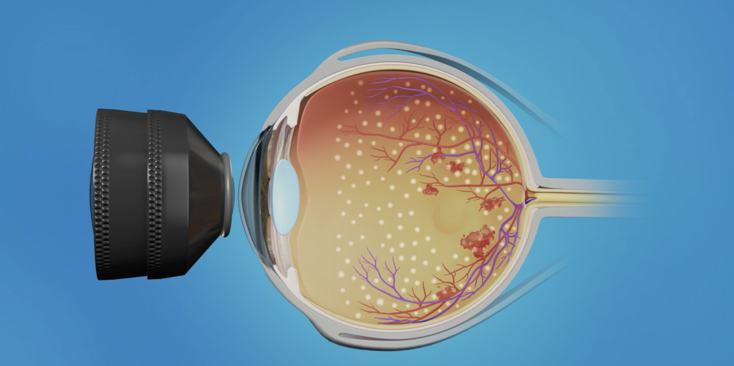
Panretinal Photocoagulation (PRP Laser) is a laser treatment used in Ophthalmology to treat abnormal blood vessels in the retina. It is commonly used for conditions like Proliferative Diabetic Retinopathy, where new, fragile blood vessels can cause bleeding or vision loss. The laser helps shrink these vessels and prevent further complications in the eye.
YAG Laser Capsulotomy (YAG Cap)
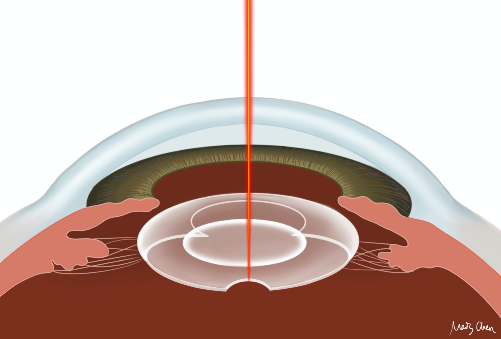
YAG capsulotomy is a laser procedure used to treat cloudiness that develops behind the artificial lens after cataract surgery. The doctor uses a laser to make a small opening in the cloudy capsule, allowing light to pass through clearly again. This helps restore clear vision and usually takes only a few minutes.
YAG Laser Peripheral Iridotomy (YAG PI)
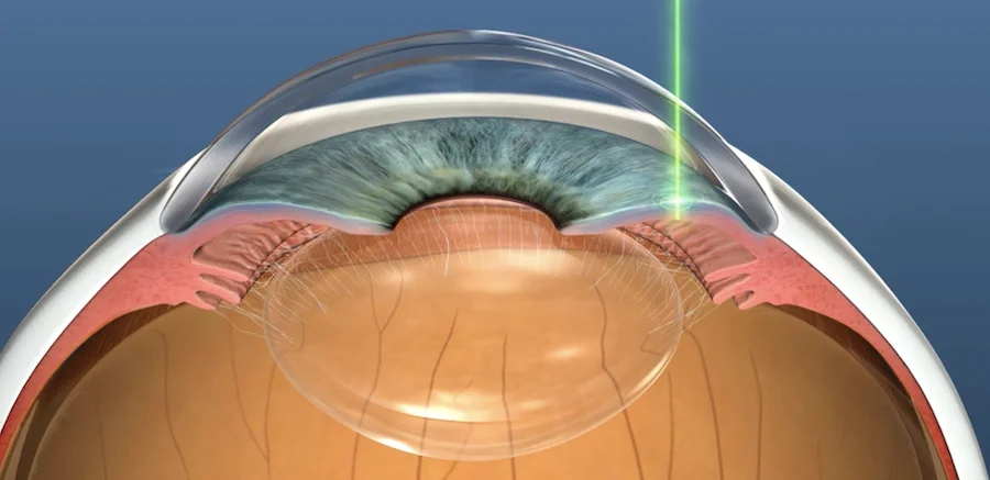
YAG peripheral iridotomy is a laser procedure used to prevent or treat certain types of glaucoma. The doctor creates a tiny hole in the colored part of the eye (iris) using a laser. This allows the fluid inside the eye to flow more freely and helps reduce eye pressure, protecting the optic nerve and vision.
Retinopexy
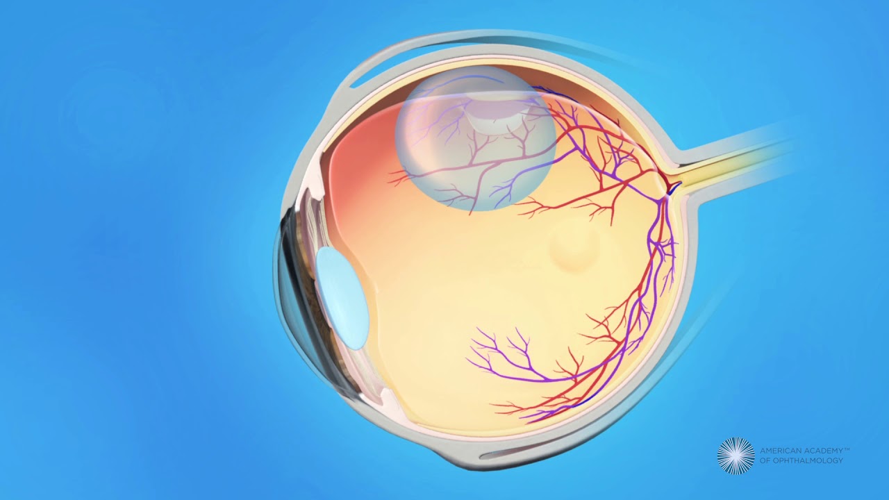
Retinopexy is an ophthalmic procedure used to create a chorioretinal adhesion around a retinal break or tear. It is performed using laser photocoagulation (laser retinopexy) or cryotherapy, which produce controlled scarring that seals the retina to the underlying retinal pigment epithelium and choroid. Retinopexy is commonly used to treat retinal tears and prevent retinal detachment, or as an adjunctive step during retinal detachment surgery to secure the retina in place.